The respiratory system is the system responsible for the exchange of gases in our bodies. As mentioned in the video above, the system is responsible for bringing air into the bloodstream and the various organs of the body, and emitting unnecessary gases from the body such as carbon dioxide (CO2).
In greater detail, the respiratory system has two roles:
Ventilation – introduction of oxygen saturated air from the external environment into the lungs, and the removal of CO2 saturated air from the lungs out into the external environment.
Exchange of gases – introduction of oxygen from the lungs into the blood, and the removal of CO2 from the blood to the lungs.
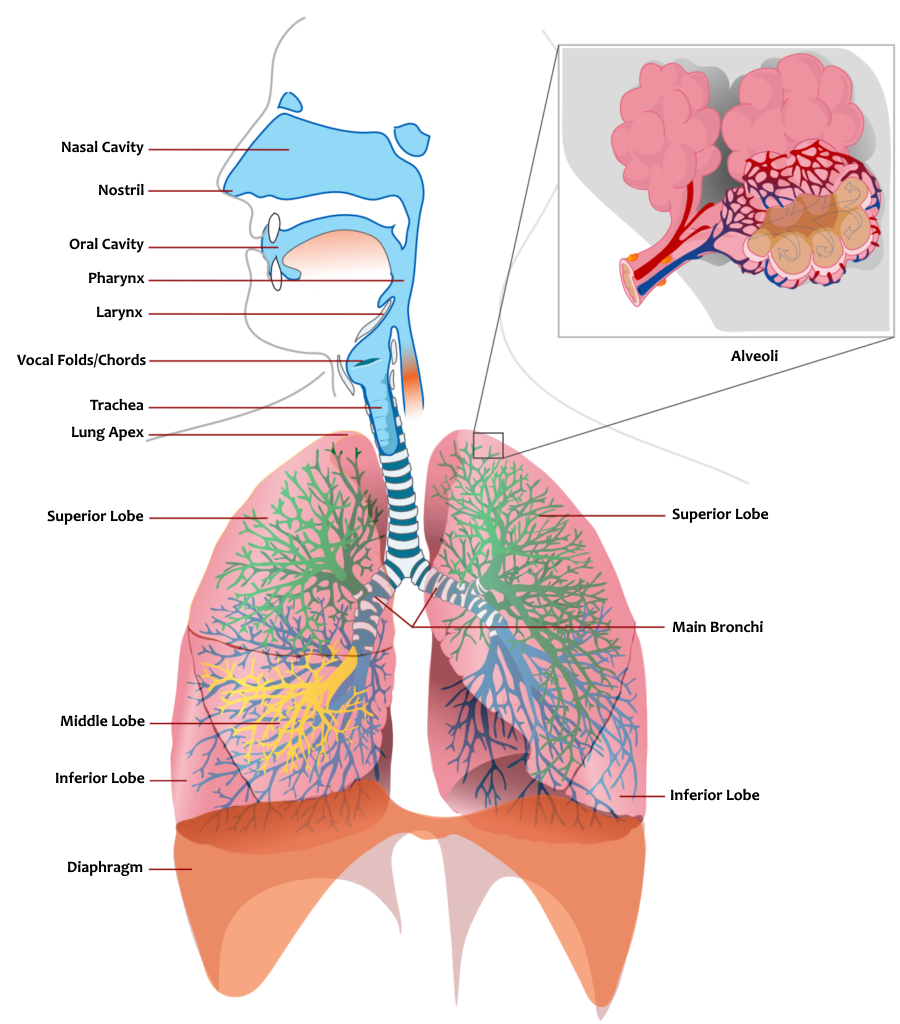
Diagram of the Respiratory System
The organs of the respiratory system are divided into upper and lower airways:
- Upper respiratory tract:
- Oral and nasal cavities
- Sinuses
- Epiglottis (the cover over the larynx)
- Vocal cords
- Trachea
- Lower respiratory tract:
- Trachea -> Bronchi -> Bronchioles -> Alveoli
- The lungs, divided into lobes: the left lung has two lobes – upper and lower, while the right lung has three lobes – upper, middle, and lower.
The lungs and ribs (which form the inside of the chest, called the thorax) are surrounded by a double membrane called the pleural membrane. The external pleura is connected to the rib cage while the internal pleura is connected to the lungs, allowing the lungs to inflate as the ribs move (since they are two separate organs). Between the pleural membranes is a small amount of liquid (10-15 ml of clear liquid) that functions as lubrication.
Air enters through the nose on its way to the trachea. In the nasal canals present in the nose, the air absorbs moisture and is heated slightly. This makes the air more effectively absorbed. These canals (nostrils) contain microscopic hairs whose job is to trap dust and other pollutants in the air, preventing them from entering the more sensitive lungs. The dirt that’s been filtered out is removed through a runny nose or mucus.
The trachea is a type of pipe that carries air from the nose to the lungs. Half of the trachea is in the throat and the other half is in the chest. Adjacent to the trachea is the esophageal tube, through which food passes into the stomach. Since the trachea and esophagus are adjacent to each other, there is a risk of food entering the trachea while swallowing. To prevent this, there is cartilage above the trachea called the epiglottis. When swallowing, the trachea rises slightly towards the epiglottis, which completely blocks it, preventing food from entering the trachea. Therefore, in situations where a person speaks while eating, there is a greater risk that the epiglottis will block the esophagus and the food will enter the trachea, causing the person to choke.
The trachea is made of rings of cartilage in the shape of a hoof so that the esophagus is not blocked from expanding when swallowing.
The trachea eventually splits into two bronchial tubes that are smaller and lead to the lungs.
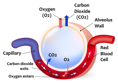
Both bronchi gradually split until they eventually become alveoli. The alveoli are microscopic cavities whose function is to transfer air to the capillaries (tiny blood vessels) that surround them, so the air can enter the bloodstream. Another role is to transfer the carbon dioxide that is absorbed from the capillaries (from the bloodstream) back into the bronchi and out of the body through exhalation. The alveoli casing has the thickness of a single cell, which allows for effective diffusion.
Diffusion is the process of dispersing material, most often caused by differences in concentration. A group of alveoli is called a lobe, with the right lung having three lobes and the left lung having two. This is because the left lung and the heart “share” space in the left thoracic region (the chest). The lobes are not dependent on each other and therefore the other lobes can still function even if one is damaged.
The thorax is a cavity that mainly contains the lungs and heart. Its boundaries are:
- From the bottom – the diaphragm, a section between the thorax and abdomen (stomach)
- From the top – the clavicle
- From the front – the sternum bone, a hard and thick bone in the center of the thorax to which the ribs are attached
- From the back – the spine, to which the ribs are attached
Most of the ribs surround the thorax from the sides and are attached to the sternum and the spine.
The exceptions are the false ribs that connect at the front to the ribs above them, and the floating ribs which, as their name implies, are not attached at the front. These ribs are connected by muscles called inter-rib muscles.
What lies between the ribs and the lungs are the pleural membranes (as explained above).
Respiration is divided into two separate processes: inhalation (inspiration) and exhalation (expiration).
This process is active, which means we are required to exercise muscles for it to happen. The diaphragm muscle, in its relaxed state, is contracted and pulled downward. Receptors located in the main arteries signal to the brain an increase in the CO2 (carbon dioxide) level in the blood. The brain sends nerve stimulation that signals the intercostal muscles to contract (and lift the thorax) and the diaphragm to contract (straightening toward the abdomen). The intercostal muscles contract and pull the ribs upward and outward. The outer pleural membrane attached to the ribs is pulled with it. The internal pleural membrane also gets pulled (the space between the membranes is a vacuum and thus the above process is possible). The lungs, which are connected to this membrane and the diaphragm, expand and produce pulmonary pressure which is lower than the external environmental pressure, causing air to enter and fill the lungs.
The concentration of oxygen entering the lungs – 21%, the concentration of carbon dioxide – 0.038%.
This process is passive, which means we do not have to exercise muscles for it to happen. In the process of exhalation, the intercostal muscles and diaphragm relax and return to their natural state, pressure exerted on the lungs causes them to contract back and expel the air outward.
The concentration of oxygen leaving the lungs – 16%, the concentration of carbon dioxide – 4%.
The normal breathing rate in an adult is between 12-20 breaths per minute. A slow breathing rate is called bradypnea and a fast breathing rate is called tachypnea.